Fractures

A bone fracture is a medical condition where the continuity of the bone is completely or incompletely broken or crack in the continuity of a bone broken. A significant percentage of bone fractures occur because of high force impact or stress. However, a fracture may also be the result of medical conditions which weaken the bones, for example osteoporosis, some cancers, or osteogenesis imperfecta/brittle bone diseases.
When discussing bones, doctors, particularly bone specialists such as orthopedic surgeons, use the term "break" much less frequently. A crack (not only a break) in the bone is also known as a fracture. Fractures can happen to any bone in the body.
The skeletal system is made up of bones and cartilage that are linked together by ligaments to form a framework for the rest of the body's tissues. The skeleton is divided into two parts:
Axial skeleton – bones that run parallel to the body's axis, such as the skull, vertebral column, and ribcage;
The appendicular skeleton consists of appendages such as the upper and lower limbs, pelvic girdle, and shoulder girdle.
Bone composition
Bone matrix has three main components:
- 25% organic matrix (osteoid);
- 50% inorganic mineral content (mineral salts);
- 25% water (Robson and Syndercombe Court, 2018).
Bone architecture is made up of two types of bone tissue: Cortical bone and cancellous bone.
Cortical bone-Also known as compact bone, this dense outer layer provides support and protection for the inner cancellous structure.
Cancellous bone/spongy bone-cancellous bone is found in the outer cortical layer. It is formed of lamellae arranged in an irregular lattice structure of trabeculae, which gives a honeycomb appearance. The large gaps between the trabeculae help make the bones lighter, and so easier to mobilize.
Types of bones
Long bones – typically longer than they are wide (such as humerus, radius, tibia, femur ), they comprise a diaphysis (shaft) and epiphyses at the distal and proximal ends, joining at the metaphysis. Most long bones are located in the appendicular skeleton and function as levers to produce movement
Short bones – small and roughly cube-shaped, these contain mainly cancellous bone, with a thin outer layer of cortical bone (such as the bones in the hands and tarsal bones in the feet)
Flat bones – thin and usually slightly curved, typically containing a thin layer of cancellous bone surrounded by cortical bone (examples include the skull, ribs and scapula). Most are located in the axial skeleton and offer protection to underlying structures
Irregular bones – bones that do not fit in other categories because they have a range of different characteristics. They are formed of cancellous bone, with an outer layer of cortical bone (for example, the vertebrae and the pelvis)
Sesamoid bones – round or oval bones (such as the patella), which develop in tendons
COMMON TYPES OF FRACTURES INCLUDE:
- Stable fracture. The broken ends of the bone line up and are barely out of place.
- Open, compound fracture. The skin may be pierced by the bone or by a blow that breaks the skin at the time of the fracture. The bone may or may not be visible in the wound.
- Transverse fracture. A straight break right across a bone. This type of fracture has a horizontal fracture line.
- Oblique fracture. This type of fracture has an angled pattern.
- Comminuted fracture. In this type of fracture, the bone shatters into three or more pieces.
Fractures can also be Classified- By quality of bone in relation to load
a) Traumatic fractures - Occurs when excessive force is applied to normal bone either directly or indirectly
b) Fatigue/Stress fractures - This occurs if bones are subjected to chronic repetitive forces, none of which alone would be enough to break the bone but which mean that the mechanical structure of the bone is gradually fatigued
c) Pathological fractures - Produced when the strength of bone is reduced by disease
SYMPTOMS
The signs and symptoms of a fracture vary according to which bone is affected, the patient’s age and general health, as well as the severity of the injury. Many fractures are excruciatingly painful, and you may be unable to move the injured area. Other common symptoms are as follows:
- Swelling and tenderness in the area of the injury
- Bruising
- Deformity — a limb may appear "out of place," or a portion of the bone may puncture the skin.
NOTE: If at all possible, do not move a person who has a broken bone until a healthcare professional arrives to assess the situation and, if necessary, apply a splint. If the patient is in a hazardous location, such as the middle of a busy road, one must sometimes act before the emergency services arrive.
PATHOPHYSIOLOGY OF FRACTURE HEALING
The pattern of healing in a given bone is influenced by;
• Rigidity of fixation of the fragments
• Closeness of their cooptation
a) Haematoma (24-48Hrs)-Injury (fracture) leads to haematoma formation from the damaged blood vessels of the periosteum, endosteum, and surrounding tissues & there is necrosis of bone immediately adjacent to the fracture.
b) Inflammation & Cellular proliferation-There is immediate release of cytokines that;
• Within hours attract an inflammatory insinuate of neutrophils and macrophages into the haematoma that debride and digest necrotic tissue and debris, including bone, on the fracture surface.
• Attract undifferentiated stem cells - probably from the periosteum & the endosteum, which migrate in & start differentiating into fibroblasts & bone-producing cells.
c) Callus formation (4-6wks)
During the reparative stage, the haematoma is gradually replaced by specialized granulation tissue with the power to form bone - callus, from both sides of the fracture. Callus is composed of fibroblasts, chondroblasts, osteoblasts and endothelial cells.
The extent to which callus forms from the periosteum, cortical bone or medulla, depends upon; the site of fracture, the degree of immobilization and the type of bone injured.
As macrophages phagocytose the haematoma and injured tissue, fibroblasts deposit a collagenous matrix, and chondroblasts deposit mucopolysaccharides in a process called endochondral bone formation. The collagenous matrix is then converted to bone as osteoblasts condense hydroxyapatite crystals on specific points on the collagen fibres, and endothelial cells form a vasculature characteristic of bone with an end result analogous to reinforced concrete. Eventually the fibrovascular callus becomes calcified - This is termed as Union.
Clinical Union - A bone is clinically united when putting load on the fracture produces no detectable movement & no pain. The fracture site will not yet be as strong as the bone around it, but it is united.
Radiological union - Occurs when the callus around the fracture can be seen to pass from one broken bone end to the other without a gap between.
d) Consolidation
This final phase, involving the replacement of woven bone (Immature bone or osteoid which is calcified callus) by lamellar bone in various shapes and arrangements, is necessary to restore the bone to optimal function. This process consolidation - involves the simultaneous meticulously coordinated removal of bone from one site (osteoclasts) and deposition in another (osteoblasts) & Ossification - the process of deposition of inorganic bone substance by osteoblasts about themselves - starts at the center of the fracture cleft, where oxygen levels may be low.
e) Remodeling-Bone is strengthened in the lines of stress & resorbed elsewhere
DIAGNOSIS
The most common way to evaluate a fracture is with x-rays, which provide clear images of bone. Your doctor will likely use an x-ray to verify the diagnosis. X-rays can show whether a bone is intact or broken. They can also show the type of fracture and exactly where it is located within the bone.
TREATMENT
This can be divided into
- REDUCTION - Reducing a fracture involves trying to return the bones to as near to their original position as possible
Methods
- Closed reduction- This is the standard initial method of reducing most common fractures. The technique is to simply grasp the fragments through the soft tissues, to disimpact them if necessary, & then to adjust them as nearly as possible to their correct position. The advantage of closed reduction is that it Minimizes damage to blood supply & soft tissues.
- Open Reduction - The fracture is exposed surgically so that the fragments can be reduced under direct vision; Fixation is usually applied to ensure that the position is maintained. A specially designed metal rod, called an intramedullary nail, provides strong fixation for this thighbone fracture.
Reduction by Mechanical traction- When large muscle contractions exert a strong displacing force, mechanical assistance may be required to draw the fragments out to the normal length of the bone. The goal may be to achieve full reduction in one sitting under anaesthesia, or to rely on gradual reduction over time. Traction for an extended period of time without anaesthesia.
- IMMOBILISATION
Indications; to relieve pain, to prevent movement that might interfere with union and to prevent displacement or angulation of the fragments - Especially fractures of the shafts of the major long bones.
The Advantages of immobilization include;
• Reduces rates of infection
• Facilitates wound care
• Promotes soft tissue healing
• Allows immobilization of the limb, particularly important in multiply injured patients
Cast Immobilization
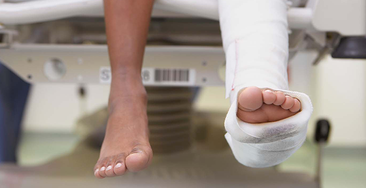
A plaster or fiberglass cast is the most common type of fracture treatment, because most broken bones can heal successfully once they have been repositioned and a cast has been applied to keep the broken ends in proper position while they heal.
Functional Cast or Brace. The cast or brace allows limited or "controlled" movement of nearby joints. This treatment is desirable for some, but not all, fractures.
Traction. Traction is usually used to align a bone or bones by a gentle, steady pulling action. Continuous traction is useful when the plane of the fracture is oblique or spiral, the elastic pull of the muscles tends to draw the distal fragment proximally, overlapping the proximal fragment.
External Fixation - In this type of operation, metal pins or screws are placed into the broken bone above and below the fracture site. The pins or screws are connected to a metal bar outside the skin. This device is a stabilizing frame that holds the bones in the proper position while they heal.
In cases where the skin and other soft tissues around the fracture are badly damaged, an external fixator may be applied until surgery can be tolerated.
Physiotherapy management of fractures
Physiotherapy should begin as soon as the fracture is immobilized. Physiotherapy will focus on the following areas during fracture healing:
- promoting health and healing
- Weight bearing is encouraged.
- Muscle strength of weakened muscles must be maintained.
- Maintaining the affected and surrounding joints' range of motion
- Pain alleviation
Physiotherapy is continued after your fracture has healed and/or your cast has been removed, or until you have regained your full level of function. The goals of physiotherapy is to Increase your weight-bearing activities, Return to normal operation, Restore muscle and joint strength and range of motion, Concentrate on sport-specific rehabilitation, Swelling reduction and Optimize the range of movement at the affected joint.
COMPLICATIONS IN FRACTURES
Early complications
a) Infection- if there is a break in the skin, as may happen with a compound fracture, bacteria can get in and infect the bone or bone marrow, which can become a persistent infection (chronic osteomyelitis).
b) Vascular injury- common vascular injuries are
Injury vessel
First rib fracture – subclavian
Shoulder dislocation- axillary
Humeral supracondylar-brachial
Elbow dislocation- brachial
Pelvic fracture- presacral and internal iliac
Femoral supracondylar fracture- femoral
Knee dislocation - popliteal
Some signs and symptoms of vascular injuries include: Paraesthesia or numbness in the toes or the fingers, Injured limb is cold & pale, or slightly cyanosed and weak or absent pulse.
- Nerve injury
- Haemarthrosis
- Visceral injury
- Gas Gangrene-This is a condition produced by Clostridium perfringens within 24Hrs of the injury characterized by myonecrosis; the patient complains of intense pain & swelling around the wound & a brownish discharge may be seen. There is little or no pyrexia but the pulse rate is increased & a characteristic smell becomes evident.
- Fracture blisters
- Plaster sores & Pressure sores
LATE COMPLICATIONS
a) Malunion-When the fragments join in an unsatisfactory position (unacceptable angulation, rotation or shortening). This may be caused by Failure to reduce a fracture adequately, Failure to immobilize while healing proceeds or Gradual collapse of comminuted or osteoporotic bone.
b) Delayed Union & Non-union -Failure of the fragments of a broken bone to knit together in time or at all
c) Disruption of bone growth – if a childhood bone fracture affects the growth plate, there is a risk that the normal development of that bone may be affected, raising the risk of a subsequent deformity.
d) Myositis ossificans - Heterotrophic bone formation or deposition of calcium in muscles with fibrosis, causing pain and swelling in muscles usually due to excessive manipulation of fractures.
e) Tendon lesions
f) Nerve compression
g) Muscle contracture
h) Joint instability/stiffness
i) Osteoarthritis
j) Fat embolism syndrome - Mainly after severe fractures of the pelvis & lower limbs, particularly those of the femur & tibia.
k) Algodystrophy - This is a syndrome comprising pain, vasomotor instability, trophic skin changes, functional impairment & osteoporosis. Follows trauma to the hand & foot & sometimes the knee, hip or shoulder.
Physiotherapy can help you return to full function faster and have a more positive rehabilitation outcome. If you are recovering from a fracture and want to maximize your rehabilitation potential with one of our specialist musculoskeletal physiotherapists, please call 0798079039 to schedule an appointment. Alternatively, you can make an appointment online right now!
Protecting your health information at every step.
Who we are
Tibabu is your go to health and medical centre, we combine our passion for love with our love for humanity. We understand that your health defines us and believe that we are God's instruments, dedicated to delivery of the best quality healthcare.
Useful Links
Our Contacts
Dereshe Towers Off Murang'a Road,
Ngara, 4107 - 00506, Nairobi