Burns

Burn injury has a devastating potential and it is one of the major health problems of the industrial world. Burns refers to any damage to the skin or other body parts caused by extreme heat, flame, contact with heated objects, or chemicals. Burns from hot liquids, steam, and fire are the most common causes of burns.
Depending on how much the burn has penetrated the skin; the burn can be categorized into four types:
1.First-degree burns (superficial burns): The damage is restricted only to the outer or topmost layer (epidermis) of the skin. The burn site is red, painful, dry, and with no blisters. Mild sunburn is an example. Long-term tissue damage is rare and often consists of an increase or decrease in the skin color.

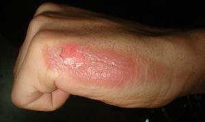
2.Second-degree burns (superficial partial thickness): The damage crosses the epidermis and reaches the layer (dermis) underneath. The burn site looks red, blistered, and may be swollen and painful.
3. Third- degree burns (deep partial thickness): The deepest layer (hypodermis) of the skin is damaged. They may go into the innermost layer of skin, the subcutaneous tissue. The burn site may look white or blackened and charred.
4. Fourth-degree burns (full-thickness burns): The skin is destroyed with damage to the underlying structures, such as nerves, tendons, and bones. There is no feeling in the area since the nerve endings are destroyed.
There are three common types of burns
1. Thermal injury
- Flame burn – Common from various causes including house fire the burn wound Can be of any depth and is usually a mix of different depths.
- Scald burn – Caused by hot fluid, about 60% of ‘burns’ in children are scalds.
- Contact burn – Caused by contacting heat sources include radiator, glass front of gas fires and irons. May cause full-thickness burns in those lost sensation, elderly, post-convulsion or toxicated by drugs and alcohol.
- Flash burn – Usually caused from ignition of a flammable substance that causes a ball of fire. Commonly results in superficial flame burn usually to the face, neck and upper limbs.
2. Electrical injury
- Low voltage – Causes by domestic electrical supplies 1,000 volts) or lightening strikes lead to significant systemic injuries and causes muscular damage that often is much greater in extent than the overlying cutaneous injury.
- High voltage-From industrial power cables (>1,000 volts) or lightening strikes lead to significant systemic injuries and causes muscular damage that often is much greater in extent than the overlying cutaneous injury.
3. Chemical injury
– Acids
– Alkali
– Organic compounds
First aid for burns
1. Ensure your own safety and Avoid self-harm.
2. Stop the burning process. – Flame source; stop, drop, cover face & roll. – Remove heat sources: hot fluid, scalding or charred clothing or remove patient from the source of injury. – In electrical injuries, disconnect the person from the source of electricity.
3. Cooling down the burn: – Cool with running tap water (8–25°C) for at least 20 minutes; Ideal temperature is 15°C. – It is believe cooling is effective up to 3 hrs. after injury, however, some studies showed no effects of cooling after 60 minutes (Nguyen et al). – Ice should not be used as it causes vasoconstriction and hypothermia. Ice can also cause burning when placed directly against the skin.
EFFECTS OF BURN ON THE BODY
• Increase vascular permeability; results in formation of oedema, compartment syndrome.
• Loss of water or hypovolaemia, electrolytes, proteins and heat (shock phase).
• Immunosuppression, which causes infection.
• Impairment of barrier function of the gut leading to translocation of bacteria.
• Systemic inflammatory response fects of burns
BURN WOUNDS HEALING PROCESS
Epidermal Healing
• The epithelial cells stop migration when they are completely in contact with other epithelial cells at the wound margins.
• This process needs to be provided by adequate nutrition and blood supply, or else the new cells will die.
Dermal Healing
• Scar formation occurs.
• Scar formation can be divided into inflammatory, Proliferative and Maturation.
INFLAMMATORY PHASE
• Inflammation begins at the time of injury, ends in about 3 to 5 days.
• Characterized by:
– Redness, edema, warmth, pain, and decreased ROM.
– blood vessel is ruptured
• Platelets aggregate, and fibrin is deposited clot formation
• Blood vessels contract decrease blood flow.
PROLIFERATIVE PHASE
• Formation of granulation tissue contains large numbers of fibroblasts macrophages, collagen, and blood vessels.
– Fibroblasts are the cells that synthesize scar tissue in random alignment
• The newly formed blood vessels bring a rich blood supply to the area and encourage further wound healing.
• Excess granulation tissue may lead to increased hypertrophic scarring and wound contraction occurs.
MATURATION PHASE
• During the maturation phase
– There is a reduction in the number of fibroblasts
– Decrease in vascularity due to a lesser metabolic demand
– Remodeling of collagen, which becomes more parallel in arrangement and forms stronger bonds.
• The ratio of collagen breakdown to production determines the type of scar that forms.
– Hypertrophic scar may result (keloid).
Acute management of burn injury
The acute management of burn injury usually takes place at emergency room or burn unit:
– Airway maintenance with cervical spine control.
– Breathing and Ventilation.
– Circulation with Hemorrhage Control.
– Disability: Neurological Status.
– Exposure with Environmental Control.
– Fluids Resuscitation.
TREATMENT OF BURN INJURIES
SURGICAL MANAGEMENT
SKIN GRAFT: a part of healthy skin is taken from a ‘donor site’, and implanted at the damaged ‘recipient site’. The skin could be from the same or another person or animal. Usually performed in a hospital under general anesthesia. Healing time depend on size and severity of injured area. Additional surgery may be required.
Aims of skin graft
• To facilitate optimal and rapid healing of the wound, minimizing deleterious consequences such as scar contracture
• Maximizing the best functional and cosmetic outcomes.
• Ameliorate the body’s systemic responses, especially the immune and metabolic systems.
Grafts and donor sites should be kept clean, moist and covered. Splinting and dressing for about 5 days following skin grafting May be reapplied to maintain proper positioning. Therapeutic exercise by physical and occupational therapy – Range of motion, strength and endurance are encouraged to resume as much independent activity as possible.
Escharotomy procedure
Escharotomy is usually done within the first 2 to 6 hours of a burn injury. Unlike fasciotomies, where incisions are made specifically to decompress tissue compartments, escharotomy incisions do not breach the deep fascial layer.
Indications
- Eschar compressing or potentially compressing tissue in or surrounding burn area Compressed tissue is identified by any of the following:
- Absent distal arterial flow as determined with a Doppler ultrasonic flow meter in the absence of systemic hypotension
- An oxygen saturation below 95% in the distal end of the extremity as shown by pulse oximetry in the absence of systemic hypoxia
- Measurement of compartment pressure > 30 mm Hg
- Impending or established respiratory compromise due to circumferential torso or neck burns
Physicians should have a high index of suspicion and a low threshold for doing escharotomy.
REHABILITATION OF BURN PATIENT
Physical and occupational therapists play an essential role in the acute management of all burn-injured patients. Realistic rehabilitative care plan and goals should be devised with multidisciplinary burn team, patient, and his /her family.
The short-term rehabilitation goal is to preserve the patient’s range of motion and functional ability.
Long-term rehabilitation goals are:
– To return patient to independent living.
– To train patients on compensating functional loss.
Acute Phase Rehabilitation
- To prevent deformity and contractures:
– Performing passive ROM.
– Splinting and antideformity positioning. •
- Reduce edema
- Establishing a long-term relationship with the patient and family members to:
– ensure compliance with therapy goals.
– increase the patient's morale for recovery
Range of Motion Exercise
• Performed twice a day.
• Exercises should be started on the first day after admission.
• Joint ranges of movement and muscle power must be documented on a chart on day one.
• Assessed and recorded on a daily basis until full active range of movement is achieved.
Splinting
• Positioning splints need not always be applied prophalactically.
– If a patient is unable to maintain proper position, and start losing ROM, splinting should be initiated.
• Positioning and splinting is an essential part of acute burn treatment regime and used for:
– Protecting joints at risk of developing contracture or deformity.
– Preserving function.
• When splinting the burn OT must be aware of the anatomy and kinesiology of the body surface to be splinted.
• Indications for Splints
– Prevention or Correction of deformity.
– Positioning - post grafting.
– Protection of exposed tendons and joints.
– Aiding in controlling edema, inflammation, or infection.
• Warning signals of bad splinting:
– Pain.
– Sensory impairment.
– Wound maceration.
Disabilities resulting from burns
If a person suffers a burn and has a physiological, psychological or anatomic problem that interferes with the normal activities of daily living, it is said that they have “impairment”. This is a medical term that directly relates to something the burn patient cannot do. Disability, on the other hand, is not a medical term but means that the person cannot meet social, personal or occupational demands placed on them because of their physical or mental limitations. If you have suffered burns, you may be unable to work and earn a living. Not only can burn-related injuries leave patients with lifelong physical disabilities but burns can also result in severe psychological and emotional distress due to scarring, which often also result in significant burdens for the patients' families and caregivers.
Complications of burns
Third-degree burns that are deep and affect a large portion of skin are very serious and can be life threatening. Even first- and second-degree burns can become infected and cause discoloration and scarring. First-degree burns do not cause scarring.
Potential complications of third-degree burns include:
- Arrhythmia, or heart rhythm disturbances, caused by an electrical burn.
- Dehydration.
- Disfiguring scars and contractures. Contractures occur when the burn scar matures, thickens, and tightens. This can prevent movement. It usually occurs when a burn occurs over a joint. A contracture is a serious complication of a burn
- Edema (excess fluid and swelling in tissues).
- Organ failure.
- Seriously low blood pressure (hypotension) that may lead to shock.
- Severe infection that may lead to amputation or sepsis. The number of organisms present on the burn wound is an important factor in infectivity. It is theorized that high temperature initially sterilizes the burn wound. However, rapid colonization by normal skin flora and existent pathogens follows.
Protecting your health information at every step.
Who we are
Tibabu is your go to health and medical centre, we combine our passion for love with our love for humanity. We understand that your health defines us and believe that we are God's instruments, dedicated to delivery of the best quality healthcare.
Useful Links
Our Contacts
Dereshe Towers Off Murang'a Road,
Ngara, 4107 - 00506, Nairobi